Digital Radiography
Introduction
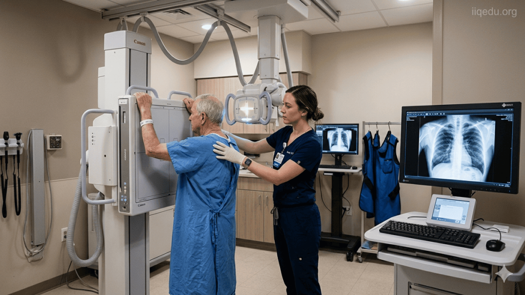
Digital radiography (DR) is an advanced form of X-ray imaging that uses digital detectors to capture radiographic images directly onto a computer system, replacing traditional film-based radiography. This technology has revolutionized medical imaging by enhancing image quality, improving workflow efficiency, and reducing radiation exposure. DR is widely used in medical, dental, veterinary, and industrial applications.
Technology and Components
Digital radiography systems are broadly classified into two main types: direct digital radiography and computed radiography (CR).
- Direct Digital Radiography (DDR):
- DDR uses flat-panel detectors made from either amorphous silicon (a-Si) or amorphous selenium (a-Se).
- X-rays interact directly with the detector to produce an electrical signal, which is then processed to form a digital image.
- The image is available almost instantly for review on a computer screen, reducing patient wait times and improving clinical efficiency.
- Computed Radiography (CR):
- CR uses photostimulable phosphor plates (PSP) instead of traditional film.
- After exposure, the plate is scanned using a laser to release stored energy as light, which is then converted into a digital image.
- While CR is slower than DDR, it allows institutions to upgrade existing X-ray equipment without fully replacing hardware.
Advantages of Digital Radiography
- Improved Image Quality:
- DR offers higher contrast resolution and better visualization of soft tissues compared to conventional film radiography.
- Post-processing tools allow manipulation of images (brightness, contrast, zoom) without additional radiation exposure.
- Reduced Radiation Exposure:
- Digital detectors are more sensitive than film, requiring lower X-ray doses to produce diagnostically useful images.
- Dose reduction is particularly significant in pediatric imaging.
- Enhanced Workflow and Storage:
- Digital images can be stored in Picture Archiving and Communication Systems (PACS), allowing immediate access across multiple locations.
- Electronic records reduce the need for physical storage space and minimize the risk of film loss or damage.
- Environmental and Cost Benefits:
- Eliminates the need for chemical processing used in film radiography, reducing environmental hazards and operational costs.
- Long-term cost savings arise from reduced film and chemical use, as well as improved efficiency.
Clinical Applications
Digital radiography is used in a wide range of clinical contexts, including:
- Chest radiography for lung evaluation and monitoring of pulmonary diseases.
- Orthopedic imaging for bone fractures, joint evaluation, and pre-surgical planning.
- Dental radiography for routine check-ups, implant planning, and endodontics.
- Pediatric imaging where dose minimization is critical.
Challenges and Limitations
- High Initial Cost: DR systems require significant upfront investment, including detectors and PACS integration.
- Technical Expertise: Proper training is required for radiographers and radiologists to optimize image acquisition and interpretation.
- Detector Sensitivity: While highly sensitive, digital detectors can be prone to artifacts from scratches, dust, or detector defects.
Future Trends
- Artificial Intelligence (AI): AI algorithms are increasingly being integrated with DR for automated detection of abnormalities, such as fractures, pulmonary nodules, or tuberculosis.
- Portable DR Units: Compact, mobile DR systems allow bedside imaging in intensive care units or remote locations.
- Dose Optimization: Advanced software continues to reduce radiation exposure while maintaining diagnostic quality.
Digital radiography continues to evolve, offering improved diagnostic capabilities, workflow efficiency, and patient safety, making it a cornerstone of modern imaging practice.
#Digital Radiography in Surat
What is Digital Radiography?
Digital radiography (DR) is a modern imaging technique that captures X-ray images using digital detectors instead of traditional photographic film. In DR, X-rays pass through the body and are detected by electronic sensors, which convert the radiation into a digital image that can be viewed, processed, and stored on a computer almost immediately. This technology is widely used in medical, dental, veterinary, and industrial imaging because it offers faster results, higher image quality, and more efficient workflows than conventional radiography.
Key Features of Digital Radiography:
- Direct Digital Capture:
- DR systems use flat-panel detectors, either based on amorphous silicon (a-Si) or amorphous selenium (a-Se), to directly convert X-ray photons into electrical signals.
- The resulting image is immediately available for review on a monitor.
- Post-Processing Capabilities:
- Digital images can be enhanced with software tools, including brightness, contrast adjustments, zoom, and edge enhancement, improving diagnostic accuracy.
- Reduced Radiation Exposure:
- Digital detectors are more sensitive than traditional film, allowing the same diagnostic quality with a lower X-ray dose, which is especially beneficial for pediatric and repeated imaging.
- Integration with Electronic Systems:
- DR images are stored digitally, often in a Picture Archiving and Communication System (PACS), allowing easy access, sharing, and long-term storage without the need for physical films.
Applications:
- Medical Imaging: Chest X-rays, bone fractures, joint evaluations, mammography, and emergency imaging.
- Dental Imaging: Routine check-ups, implant planning, and endodontic procedures.
- Veterinary and Industrial Uses: Animal health diagnostics and non-destructive testing of materials.
Benefits of Digital Radiography:
- Faster image acquisition and immediate availability.
- Improved image quality and diagnostic accuracy.
- Reduced environmental impact due to elimination of film processing chemicals.
- Enhanced workflow efficiency and record management.
#Digital Radiography in Delhi
Who is Digital Radiography required?
Digital radiography (DR) is a diagnostic tool used in a wide range of clinical, dental, and industrial settings. Its requirement is determined by the need for high-quality imaging, quick results, and minimal radiation exposure. DR is not limited to specific patients; instead, it is indicated whenever X-ray imaging is necessary and when the benefits of digital imaging outweigh traditional film-based radiography.
1. Medical Patients
DR is commonly required in hospitals, clinics, and emergency care for patients who need:
- Chest Imaging:
- Suspected pneumonia, tuberculosis, lung cancer, or COVID-19 complications.
- Monitoring chronic conditions like asthma or chronic obstructive pulmonary disease (COPD).
- Orthopedic Assessment:
- Evaluation of bone fractures, joint dislocations, arthritis, or spinal injuries.
- Preoperative and postoperative monitoring for surgical planning and recovery.
- Abdominal and Gastrointestinal Imaging:
- Detection of intestinal obstructions, kidney stones, or swallowed foreign objects.
- Pediatric Patients:
- DR is preferred due to lower radiation exposure, faster image acquisition, and the ability to reduce repeat scans.
- Trauma and Emergency Patients:
- Rapid imaging in accidents, falls, or trauma cases where timely diagnosis is critical.
2. Dental Patients
- DR is widely used in dentistry for:
- Routine check-ups to detect cavities or periodontal disease.
- Endodontic procedures like root canals.
- Implant planning or orthodontic assessments.
- Reduces radiation dose compared to traditional dental X-ray film.
3. Veterinary Patients
- Animals needing internal imaging for fractures, dental issues, or disease evaluation benefit from DR.
- Rapid imaging improves care in emergency situations for pets or livestock.
4. Industrial and Research Applications
- DR is required in non-destructive testing (NDT), including:
- Inspecting welds, pipelines, or structural components.
- Quality assurance in manufacturing.
- Security screening for critical infrastructure.
5. High-Risk Groups
Certain patient groups particularly benefit from DR due to its low-dose radiation and fast imaging:
- Children – more sensitive to radiation.
- Pregnant women – only when imaging is unavoidable, with DR reducing exposure.
- Patients requiring frequent imaging – chronic conditions needing repeated X-rays.
Summary
Digital radiography is required whenever precise, rapid, and safe imaging is needed. It is especially beneficial for:
- Patients needing frequent or repeat imaging.
- Pediatric and high-risk populations.
- Emergency, orthopedic, or trauma cases.
- Dental assessments and implant planning.
- Industrial inspections where digital detection is more efficient than traditional methods.
#Digital Radiography in Pune
When is Digital Radiography required?
Digital radiography (DR) is required whenever X-ray imaging is necessary for diagnosis, treatment planning, or monitoring, particularly when efficiency, image quality, and reduced radiation exposure are priorities. The decision to use DR depends on the patient’s condition, the type of examination needed, and the clinical setting.
1. Trauma and Emergency Situations
- Acute injuries such as fractures, dislocations, or internal trauma require immediate imaging.
- DR provides rapid image acquisition for emergency evaluation, allowing faster clinical decisions.
- Examples include: car accidents, falls, or sports injuries.
2. Routine Diagnostic Imaging
- Chest X-rays for conditions such as pneumonia, tuberculosis, lung cancer, or chronic respiratory diseases.
- Abdominal and gastrointestinal imaging to detect obstructions, kidney stones, or swallowed foreign objects.
- Bone and joint evaluation for suspected fractures, arthritis, or degenerative changes.
3. Pediatric and High-Risk Populations
- DR is preferred when radiation dose reduction is essential, such as in children, pregnant women (with precautions), and patients requiring frequent imaging.
- Pediatric imaging benefits from DR’s speed and sensitivity, minimizing the need for repeat exposures.
4. Preoperative and Postoperative Assessment
- Orthopedic surgeries: DR is used for preoperative planning, confirming bone alignment, or checking surgical implants postoperatively.
- Dental procedures: Preoperative imaging is necessary for implants, root canal therapy, and orthodontic planning.
5. Chronic Disease Monitoring
- Patients with chronic conditions such as osteoporosis, scoliosis, or pulmonary disease require periodic imaging.
- DR reduces cumulative radiation exposure while maintaining image quality over multiple scans.
6. Industrial and Non-Medical Applications
- In non-destructive testing (NDT), DR is required to inspect pipelines, welds, or structural components without damaging them.
- DR provides high-resolution images quickly, improving workflow in quality control and safety inspections.
7. Situations Requiring Immediate Digital Access
- When images need to be stored, shared, or reviewed remotely using PACS or teleradiology systems.
- Useful for multi-specialty consultations, large hospitals, or clinics where instant access improves patient care.
Summary
Digital radiography is required in situations that demand:
- Rapid imaging and diagnosis (emergencies, trauma).
- Reduced radiation exposure (pediatrics, repeated imaging).
- High-quality images for soft tissue or subtle bone assessment.
- Pre- and post-procedural evaluation (surgical planning, dental implants).
- Integration with digital systems for storage, sharing, and remote access.
- Industrial inspections where non-destructive and precise imaging is necessary.
#Digital Radiography in Kolkata
Where is Digital Radiography required?
Digital radiography (DR) is required in a wide range of locations where X-ray imaging is essential for diagnosis, treatment, monitoring, or industrial inspection. Its use is dictated by the need for rapid, high-quality imaging with minimal radiation exposure and efficient digital workflow.
1. Hospitals and Medical Centers
- Emergency Departments:
DR is critical in trauma units for rapid assessment of fractures, dislocations, chest injuries, and internal trauma. - Radiology Departments:
Used for routine diagnostic imaging including chest X-rays, abdominal scans, skeletal evaluations, and mammography. - Intensive Care Units (ICUs):
Portable DR units allow bedside imaging for critically ill patients who cannot be transported. - Surgical Units:
Preoperative and postoperative imaging for orthopedic, dental, or maxillofacial surgeries.
2. Dental Clinics and Oral Health Facilities
- DR is standard in dental imaging for:
- Routine dental examinations to detect cavities, impacted teeth, or periodontal disease.
- Endodontic procedures such as root canals.
- Implant planning and orthodontics, allowing accurate visualization with minimal radiation.
3. Outpatient Clinics and Diagnostic Centers
- Primary care centers and specialized diagnostic imaging centers use DR for:
- Routine screening X-rays (chest, spine, and extremities).
- Follow-up imaging for chronic conditions.
4. Veterinary Clinics
- DR is widely used in veterinary medicine for:
- Diagnosing bone fractures, joint disorders, or internal organ conditions in pets or livestock.
- Dental imaging in animals, similar to human dentistry.
5. Industrial and Non-Medical Applications
- Non-Destructive Testing (NDT):
DR is used to inspect welds, pipelines, and structural components without causing damage. - Manufacturing Quality Control:
Detects defects or inconsistencies in machinery parts, castings, and critical infrastructure. - Security Screening:
Airports and sensitive facilities use DR technology to detect hidden objects or threats.
6. Mobile and Remote Locations
- Portable DR units allow imaging outside traditional facilities, including:
- Field hospitals and disaster relief zones.
- Remote communities lacking permanent radiology infrastructure.
- Military or emergency response units.
Summary
Digital radiography is required wherever fast, accurate, and safe imaging is needed, including:
- Hospitals (emergency, ICU, surgical units)
- Dental and orthodontic clinics
- Outpatient diagnostic centers
- Veterinary facilities
- Industrial and non-destructive testing sites
- Mobile, remote, or field-based healthcare setups
Its versatility across clinical and non-clinical environments makes DR an essential tool for modern imaging.
#Digital Radiography in Banglore
How is Digital Radiography required?
Digital radiography (DR) is required through a structured clinical or industrial workflow that ensures accurate imaging, patient safety, and optimal use of technology. The “how” encompasses selection of patients or objects, imaging protocols, equipment setup, and image management, along with integration into healthcare or industrial systems.
1. Clinical Requirement Process
DR is required in clinical practice when a physician, dentist, or healthcare provider identifies a need for imaging to diagnose, monitor, or guide treatment. This process typically involves:
- Clinical Assessment:
- Evaluation of symptoms, trauma, or chronic conditions.
- Determining if radiographic imaging is necessary based on clinical guidelines (e.g., American College of Radiology (ACR) appropriateness criteria).
- Selection of Imaging Modality:
- Decision between digital radiography, computed radiography (CR), or conventional X-ray.
- DR is preferred when fast results, digital storage, or post-processing adjustments are important.
- Patient Preparation:
- Positioning the patient correctly to target the area of interest.
- Removal of any objects that may interfere with image quality, such as jewelry or clothing with metal.
- Explaining the procedure and ensuring patient consent where required.
2. Technical Implementation
DR requires careful use of equipment and protocols to produce high-quality images:
- Equipment Setup:
- Using flat-panel digital detectors or photostimulable phosphor plates (PSP) for image capture.
- Calibration of X-ray machine parameters (kVp, mA, exposure time) based on patient size, anatomy, and diagnostic requirements.
- Image Acquisition:
- The patient or object is exposed to X-rays, which are captured directly by the digital detector.
- Modern DR systems produce images immediately on a computer monitor, enabling real-time review.
- Post-Processing:
- Digital images can be enhanced using software for contrast adjustment, edge enhancement, zoom, and noise reduction.
- Radiologists or clinicians review images for diagnostic accuracy, and adjustments can be made without repeating the exposure.
- Storage and Sharing:
- Images are stored in a Picture Archiving and Communication System (PACS).
- Allows secure sharing between specialists, remote consultation, and integration with electronic health records (EHR).
3. Industrial and Non-Medical Requirement
In non-clinical settings, DR is required to inspect materials or structures using similar protocols adapted for objects instead of patients:
- Object Selection:
- Choose materials or components requiring inspection for defects or integrity issues.
- Positioning and Exposure:
- Place the object between the X-ray source and digital detector.
- Adjust exposure parameters based on material density and thickness.
- Image Analysis:
- Inspect captured images for internal defects, cracks, voids, or weld quality.
- Images can be digitally archived or integrated into quality control databases.
4. When DR Is Required
DR is required under the following conditions:
- When rapid imaging is needed for clinical decisions (e.g., trauma, emergency cases).
- When reduced radiation exposure is necessary (e.g., children, frequent imaging).
- When digital storage, sharing, or remote consultation is required.
- For industrial inspections needing high-resolution digital images without destructive testing.
Summary
Digital radiography is required by clinical judgment or industrial need and implemented through a structured workflow:
- Assess the need based on symptoms, conditions, or inspection requirements.
- Prepare the patient or object and select the appropriate imaging protocol.
- Acquire the image using a digital detector system.
- Post-process, review, and store images digitally.
- Use images for diagnosis, treatment planning, monitoring, or quality assurance.
By following these steps, DR ensures high-quality imaging, efficiency, safety, and long-term digital record management.
#Digital Radiography in Chennai
Case Study of Digital Radiography
The following case studies illustrate how digital radiography (DR) has been implemented in real-world settings, demonstrating improvements in clinical workflow, diagnostic efficiency, and operational performance compared with traditional film‑based radiography. These examples are drawn from peer‑reviewed research and documented clinical experience.
1. Clinical Workflow and Efficiency at a Major Hospital
A study conducted at The Cleveland Clinic Foundation in Cleveland, Ohio, evaluated the impact of transitioning from traditional film radiography to a digital radiography system with and without integration into a Radiology Information System (RIS). Researchers compared the time required for examinations under three conditions — conventional film, DR without RIS integration, and DR with RIS integration.
Key Findings:
- DR significantly reduced the time needed to complete an X‑ray examination compared with traditional film systems.
- Real‑time quality control was possible because images were available instantly at the modality, eliminating the need for film processing and centralized review.
- Integration with RIS further reduced total examination time by facilitating automatic population of patient demographic information and streamlined workflow.
- The study demonstrated that DR improves overall productivity and can enhance patient throughput in a busy radiology department.
This case highlights how digital systems accelerate imaging workflows, reduce manual tasks, and support integration with health information systems, leading to measurable time savings and improved efficiency.
2. Conversion from Analog to Digital in Radiology Practice
At Christie Hospital NHS Trust (Manchester, UK), a transition from fully analog X‑ray procedures to a digital radiography environment was studied, focusing on chest radiography workflow. Chest exams were selected due to their prevalence and importance in diagnostic imaging.
Results:
- Average examination time was reduced from approximately 9.24 minutes under analog radiography to 5.28 minutes using digital radiography.
- The reduction in modality‑specific tasks (e.g., handling film cassettes, development time) contributed significantly to increased throughput and productivity.
- Room utilization efficiency improved as a result of the faster imaging process.
This case demonstrates that DR conversion can have a substantial impact in clinical settings where demand for X‑ray services is high and workflow optimization is critical.
3. Quality Control in Digital Radiography Systems
A study published in the Journal of Applied Clinical Medical Physics examined quality control practices in a DR system over a prolonged period, comparing traditional quality assurance methods with a more contemporary approach (termed Medical Physics 3.0).
Insights:
- Conventional annual testing and automated manufacturer quality assurance procedures sometimes failed to identify performance degradation in DR detectors.
- Advanced analysis of signal‑to‑noise ratios and clinical image quality metrics over many years revealed issues that routine tests missed.
- Adjustments based on this deeper analysis improved detector performance and maintained high‑quality breast and body imaging over time.
Although this example focuses more on quality control than operational workflow, it underscores the importance of rigorous ongoing evaluation in DR practice to sustain diagnostic accuracy and patient safety.
4. Case Example: Veterinary Application and Economic Benefits
In a veterinary practice (Neel Veterinary Hospital, Oklahoma City), DR adoption demonstrated not only clinical benefits but also operational and financial advantages:
- The practice transitioned to a digital system and eliminated costs associated with film processing and storage.
- Repeat radiographs due to processing errors decreased, reducing radiation exposure to staff and animals.
- The space previously used for film archive was repurposed, and annual operational savings were significant.
- Increased radiography throughput and the ability to offer rapid imaging led to higher gross revenue within the first year of DR system implementation.
This case exemplifies how digital radiography can improve practice efficiency and profitability beyond human healthcare settings.
5. Supporting Evidence from Global Radiology Practice
Other studies, such as those evaluating radiography workflows supported by Picture Archiving and Communication Systems (PACS), have consistently found that digital imaging improves quality and reduces repeat scans, enhances image retrieval, and streamlines radiographer performance. These benefits contribute to a higher standard of clinical service delivery and patient care across diverse settings.
Summary of Case Study Insights
Across multiple clinical and operational environments, digital radiography has been shown to:
- Reduce examination and processing time through immediate digital capture and elimination of film development steps.
- Improve workflow efficiency by integrating with RIS/PACS and automating routine tasks.
- Enhance diagnostic quality and long‑term quality control when combined with advanced evaluation methods.
- Lower operational costs by reducing film use, storage needs, and repeat imaging.
- Increase patient throughput, enabling radiology departments to serve more patients effectively.
#Digital Radiography in Ahemdabad
White Paper of Digital Radiography
1. Executive Summary
Digital radiography (DR) represents a transformational evolution in diagnostic imaging, replacing analog film‑based X‑ray systems with digital detectors that produce high‑resolution images instantly. DR has become the standard of care in medical, dental, veterinary, and industrial applications due to its superior image quality, workflow efficiency, dose reduction, and integration with digital information systems. This white paper examines the principles, technology, clinical value, implementation strategies, challenges, economic considerations, and future directions of digital radiography.
2. Introduction
Radiography has been an essential diagnostic modality since the discovery of X‑rays in 1895. Traditional film screen radiography required physical film processing, chemical usage, and manual storage, limiting throughput and flexibility. Digital radiography, introduced in the late 20th century, uses electronic detectors and digital data processing to overcome these limitations. DR enables near‑instant image acquisition, advanced image optimization, and seamless integration with healthcare information systems.
Key characteristics of DR include:
- Direct digital capture using solid state detectors.
- Post‑acquisition image processing without repeat exposures.
- Electronic storage and distribution via Picture Archiving and Communication Systems (PACS) and Health Information Exchanges (HIEs).
For foundational understanding of DR technology and clinical application, consult Bushberg et al., The Essential Physics of Medical Imaging, 3rd Edition and the International Atomic Energy Agency’s technical guidance on digital radiography.
External references:
- Bushberg JT, Seibert JA, Leidholdt EM, Boone JM. The Essential Physics of Medical Imaging. Lippincott Williams & Wilkins, 2012.
https://www.wiley.com/en-us/The+Essential+Physics+of+Medical+Imaging%2C+3rd+Edition‑p‑9781451199972 - International Atomic Energy Agency. Digital Radiography: Fundamentals and Clinical Applications. 2017.
https://www.iaea.org/publications/10547/digital‑radiography
3. Technology Overview
3.1 Detector Types
Digital radiography systems fall into two main categories:
Direct Digital Radiography (DDR):
- Uses flat‑panel detectors based on amorphous selenium (a‑Se) or amorphous silicon (a‑Si) with a scintillator.
- X‑rays are converted directly (a‑Se) or indirectly (a‑Si + scintillator) into electrical signals.
- Offers real‑time imaging with minimal delay.
Computed Radiography (CR):
- Uses photostimulable phosphor plates (PSP).
- After exposure, the plate is scanned with a laser to extract image data.
- Often retrofits existing X‑ray units, providing a cost‑effective upgrade path.
3.2 Image Acquisition and Processing
Once the detector captures the X‑ray signal, digital image processing algorithms enhance diagnostic contrast, reduce noise, and allow manipulation of image parameters (e.g., brightness, contrast, edge enhancement) without additional radiation exposure.
3.3 System Integration
DR systems typically integrate with:
- PACS for archiving and retrieval.
- Radiology Information Systems (RIS) for scheduling, workflow tracking, and reporting.
- Electronic Health Records (EHR) for longitudinal image accessibility.
4. Clinical Applications
Digital radiography is applied wherever diagnostic imaging is needed:
4.1 Medical Imaging
- Chest Radiography: Lung pathology, tuberculosis screening, cardiac silhouette evaluation.
- Skeletal Imaging: Fracture assessment, joint evaluation, degenerative disease monitoring.
- Trauma Care: Rapid decision support in emergency departments.
- Mobile and Bedside Imaging: Intensive care units and field settings.
4.2 Dental and Maxillofacial Imaging
Used for routine diagnostics, orthodontic planning, implant placement, and endodontic evaluation, with significantly lower dose than conventional dental film.
4.3 Veterinary Medicine
DR supports skeletal, thoracic, and dental examinations in animals, improving throughput and minimizing stress during imaging.
4.4 Industrial and Non‑Medical Use
- Non‑Destructive Testing (NDT): Identification of internal defects in welds, castings, and infrastructure components.
- Security Screening: Detection of concealed threats in security checkpoints.
5. Benefits of Digital Radiography
5.1 Diagnostic Quality
DR systems deliver higher spatial and contrast resolution than film, facilitating detection of subtle pathology. Digital enhancement tools improve the visibility of soft tissue structures and fine bone detail.
5.2 Radiation Dose Reduction
Detector sensitivity allows diagnostic quality images at lower doses than analog systems, contributing to safer imaging protocols, especially for pediatric and repeat studies.
5.3 Workflow Efficiency
Instant image availability eliminates film processing, reduces retakes, and accelerates patient throughput. Integration with RIS and PACS further streamlines operations.
5.4 Cost and Environmental Impact
DR reduces expenditures on film, chemicals, and storage space. The elimination of chemical processing is environmentally beneficial.
6. Implementation Considerations
6.1 Clinical Workflow Integration
A successful DR implementation includes:
- Structured protocols for patient positioning and exposure parameters.
- Training programs for technologists in digital image optimization and quality assurance.
- Defined pathways for image review, reporting, and archiving.
6.2 Technical Infrastructure
Requirements include:
- Robust networking for rapid image transfer.
- Adequate data storage and backup mechanisms.
- Interoperability with existing EHR, RIS, and PACS.
6.3 Quality Assurance
Ongoing quality control is essential to maintain detector performance and image fidelity. Advanced QA protocols may include periodic calibration, artifact detection, and image quality benchmarking. See peer‑reviewed analysis on DR quality control in the Journal of Applied Clinical Medical Physics for guidance on optimizing QA practices beyond routine testing.
Reference:
Carver DE et al. Medical Physics 3.0 vs 1.0: A Case Study in Digital Radiography Quality Control. J Appl Clin Med Phys. 2018.
https://pubmed.ncbi.nlm.nih.gov/30117273/
7. Economic Impact
7.1 Cost‑Benefit Analysis
Initial DR system acquisition costs are higher than film systems, but operational savings are realized through reduced consumables, improved efficiency, and decreased repeat imaging. Facilities report increased throughput and enhanced revenue when DR reduces bottlenecks in high‑volume departments.
7.2 Return on Investment (ROI)
Examples from veterinary and medical practices demonstrate ROI through operational cost savings and increased capacity utilization. Economic evaluations consistently show favorable long‑term returns when DR replaces analog systems.
8. Challenges and Limitations
8.1 Capital Expenditure
High upfront costs for detectors, network infrastructure, and PACS integration remain a barrier for some facilities.
8.2 Technical Expertise
DR systems require trained personnel for optimal performance, including radiologic technologists and IT support for system maintenance.
8.3 Image Artifacts
Digital detectors can exhibit artifacts from detector defects, dust, or calibration issues, necessitating vigilant quality control.
9. Future Directions
9.1 Artificial Intelligence (AI) and Assistive Analytics
AI algorithms are increasingly being incorporated to assist in detection of pathology (e.g., lung nodules, fractures) and to prioritize urgent studies in radiology workflows.
9.2 Cloud‑Based Imaging Solutions
Cloud integration offers scalable storage, remote access, and distributed diagnostics, supporting tele‑radiology and multi‑site practices.
9.3 Portable and Point‑of‑Care Systems
Miniaturized DR units enable bedside imaging and rapid diagnostics in emergency, intensive care, and remote settings.
10. Conclusion
Digital radiography is a cornerstone of modern imaging practice, combining high diagnostic quality, efficient workflow, lower radiation dose, and digital interoperability. Its implementation across healthcare environments improves patient care, accelerates clinical decision‑making, and supports advanced diagnostic services. Industrial adoption further demonstrates DR’s versatility beyond medical use.
11. References (Selected)
- Bushberg JT, Seibert JA, Leidholdt EM, Boone JM. The Essential Physics of Medical Imaging. Lippincott Williams & Wilkins, 2012.
https://www.wiley.com/en-us/The+Essential+Physics+of+Medical+Imaging%2C+3rd+Edition‑p‑9781451199972 - International Atomic Energy Agency. Digital Radiography: Fundamentals and Clinical Applications. 2017.
https://www.iaea.org/publications/10547/digital‑radiography - Seeram E. Digital Radiography: An Overview. Elsevier, 2015.
https://www.elsevier.com/books/digital‑radiography/seeram/978‑0‑323‑27294‑4 - Carver DE et al. Medical Physics 3.0 vs 1.0: A Case Study in Digital Radiography Quality Control. J Appl Clin Med Phys. 2018.
https://pubmed.ncbi.nlm.nih.gov/30117273/ - Dackiewicz D et al. “Impact of digital radiography on clinical workflow and patient satisfaction.” Journal of Digital Imaging.
https://pmc.ncbi.nlm.nih.gov/articles/PMC3453299/
#Digital Radiography in Hyderabad
Industry Application of Digital Radiography
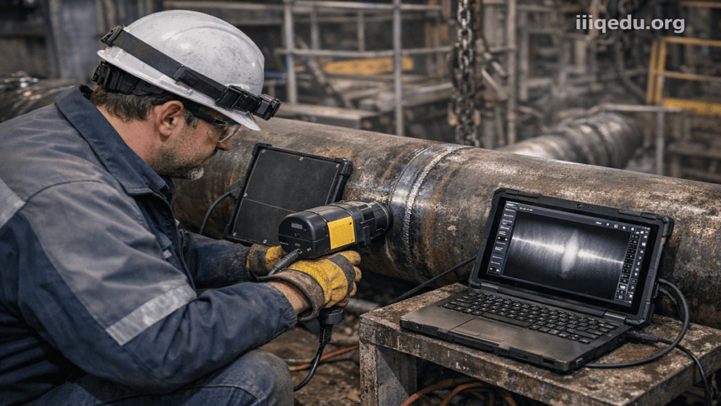
Digital radiography (DR) is widely used beyond healthcare, particularly in industrial sectors where non-destructive evaluation, quality assurance, and safety inspection are critical. By replacing traditional film-based radiography, DR enables faster imaging, improved defect detection, and seamless digital integration into industrial workflows.
1. Non-Destructive Testing (NDT)
The most significant industrial application of digital radiography is in Non-Destructive Testing (NDT), where materials and structures are inspected without causing damage.
Applications:
- Inspection of weld joints in pipelines, pressure vessels, and structural frameworks.
- Detection of internal defects such as cracks, voids, porosity, and inclusions.
- Evaluation of corrosion and material degradation in critical infrastructure.
Industries Using NDT:
- Oil and gas
- Power generation
- Construction and infrastructure
- Aerospace and defense
Advantages:
- Immediate image availability for analysis
- Higher defect detection sensitivity compared to film
- Digital storage for traceability and compliance
2. Manufacturing and Quality Control
DR plays a crucial role in ensuring product reliability and maintaining manufacturing standards.
Applications:
- Inspection of castings and molded components for internal defects.
- Verification of assembly integrity in complex machinery.
- Detection of flaws in electronics, circuit boards, and micro-components.
Benefits:
- Reduces production defects and recalls
- Enables real-time quality monitoring
- Integrates with automated production systems
3. Aerospace Industry
The aerospace sector requires extremely high standards of safety and precision.
Applications:
- Inspection of aircraft components, including turbine blades and fuselage structures.
- Detection of fatigue cracks in critical load-bearing parts.
- Evaluation of composite materials used in modern aircraft design.
Importance:
- Prevents catastrophic failures
- Ensures compliance with aviation safety regulations
- Supports lifecycle maintenance of aircraft
4. Oil and Gas Industry
In the oil and gas sector, DR is essential for maintaining the integrity of pipelines and equipment.
Applications:
- Inspection of pipeline welds and joints.
- Monitoring corrosion, erosion, and structural damage.
- Evaluation of offshore and onshore drilling equipment.
Advantages:
- Portable DR systems enable field inspections in remote locations.
- Reduces downtime by enabling rapid diagnosis and repair decisions.
5. Automotive Industry
DR is widely used in automotive manufacturing for quality assurance and safety.
Applications:
- Inspection of engine components, gear systems, and chassis structures.
- Detection of defects in welds and assembled parts.
- Testing of battery systems in electric vehicles.
Benefits:
- Improves vehicle safety and reliability
- Supports compliance with industry standards
- Enhances production efficiency
6. Security and Defense
Digital radiography is applied in security systems for threat detection and surveillance.
Applications:
- Airport baggage scanning and cargo inspection.
- Detection of weapons, explosives, or contraband in luggage and shipments.
- Military applications for equipment inspection and maintenance.
Advantages:
- High-speed screening with digital analysis
- Enhanced detection accuracy
- Integration with automated threat recognition systems
7. Cultural Heritage and Archaeology
DR is increasingly used in preservation and research of historical artifacts.
Applications:
- Examination of ancient artifacts, paintings, and sculptures without damage.
- Detection of hidden structures or repairs in artworks.
- Study of mummies and fossils for scientific research.
Benefits:
- Non-invasive analysis
- Preservation of fragile objects
- Detailed internal visualization
8. Electronics and Semiconductor Industry
Miniaturization in electronics requires precise inspection methods.
Applications:
- Inspection of printed circuit boards (PCBs).
- Detection of micro-cracks, soldering defects, and voids.
- Quality control in semiconductor manufacturing.
Advantages:
- High-resolution imaging of microscopic structures
- Early detection of faults improves product reliability
9. Food and Pharmaceutical Industry
DR is used for safety and quality control in consumable products.
Applications:
- Detection of foreign objects (metal, glass, bone fragments) in food products.
- Inspection of packaging integrity.
- Verification of tablet consistency in pharmaceuticals.
Benefits:
- Ensures consumer safety
- Meets regulatory standards
- Reduces product recalls
Summary
Digital radiography has become an essential tool across industries due to its ability to provide:
- Non-destructive internal inspection
- High-resolution and accurate defect detection
- Faster imaging and real-time analysis
- Digital storage and traceability
- Improved safety and quality assurance
Its integration into industrial systems supports efficient production, regulatory compliance, and risk reduction, making DR a cornerstone technology in modern industrial operations.
References and Further Reading
- International Atomic Energy Agency (IAEA). Industrial Radiography Manual.
https://www.iaea.org/publications - American Society for Nondestructive Testing – Resources on industrial radiography and NDT standards
https://www.asnt.org - Seeram, E. Digital Radiography: Physical Principles and Quality Control. Elsevier, 2019.
https://www.elsevier.com - Bushberg JT et al. The Essential Physics of Medical Imaging, 3rd Edition.
https://www.wiley.com
#Digital Radiography in Mumbai
Ask FAQs
What is Digital Radiography?
Digital radiography (DR) is an advanced imaging technique that uses digital detectors instead of traditional photographic film to capture X-ray images. These images are immediately available on a computer, allowing for faster diagnosis, enhanced image quality, and easy storage and sharing through digital systems.
What are the main advantages of Digital Radiography?
Digital radiography offers several advantages over conventional film-based radiography, including:
Faster image acquisition and immediate viewing
Reduced radiation exposure to patients
Enhanced image quality with post-processing capabilities
Elimination of film and chemical processing
Easy storage and sharing through digital platforms such as PACS
Where is Digital Radiography commonly used?
Digital radiography is widely used in multiple fields, including:
Hospitals and diagnostic imaging centers
Dental clinics for oral imaging
Veterinary practices for animal diagnostics
Industrial sectors for non-destructive testing and quality control
Security systems such as airport baggage screening
Is Digital Radiography safe?
Yes, digital radiography is generally considered safe when used appropriately. It uses lower radiation doses compared to traditional X-ray systems due to more sensitive detectors. Additionally, strict safety guidelines and radiation protection measures are followed to minimize exposure for both patients and operators.
How does Digital Radiography improve workflow efficiency?
Digital radiography improves workflow by eliminating the need for film processing and enabling instant image availability. Images can be reviewed, enhanced, and shared electronically, reducing examination time, minimizing repeat scans, and allowing healthcare professionals to make quicker and more accurate decisions.
Source: radiology technical
Table of Contents
Disclaimer:
This information is provided for educational and informational purposes only and should not be considered as professional medical or technical advice. Always consult a qualified professional or specialist for diagnosis, treatment, or specific application guidance.